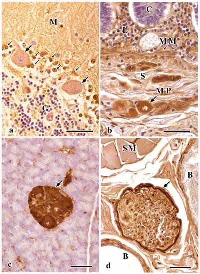
 Figure 1. Immunohistochemical localization by light microscopy of the galectin-1 in human nervous tissues. Sections were stained with anti-galectin-1 antibody and HRP-labeled goat anti-rabbit IgG. Nuclei were counterstained with hematoxylin.
a) Cerebellum. Neurons (small arrows) interspersed between Purkinje cells are stained. M: molecular layer, G: granular layer. Large arrows indicate the Purkinje cells.
b) Ileum. Meissner's plexus (MP) is stained. C: crypt, L: lamina propria, MM: muscularis mucosa, S: submucosa.
c) Pancreas. Ganglion (arrow) in the lobule of pancreas is stained intensely.
d) Omohyoid muscle. Perineurium (arrow) and axon (arrowheads) in the nerve fiber bundle are intensely stained. SM: striated muscle, B: blood vessel. Bars, 50 um.
GO BACK TO THE TEXT