Links
Links Contact Us
 Japanese
 Links
Links Contact Us
 Japanese
Research
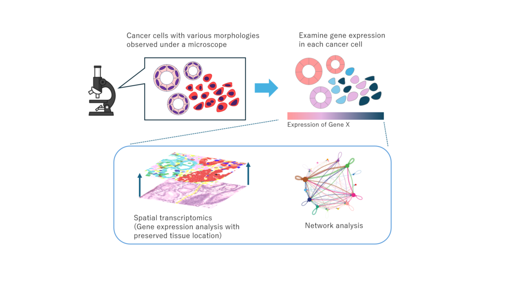
Investigation of Intratumoral Heterogeneity in Pancreatobiliary Cancer
Pancreatobiliary cancers are often already advanced at the time of diagnosis and remain among the most difficult solid tumors to treat. Even among patients diagnosed with the same type of pancreatobiliary cancer, responses to treatment and the speed of disease progression can vary greatly. One reason for this difference is thought to be intratumoral heterogeneity, meaning that cancer cell populations with different characteristics coexist within a single tumor. Previous studies have shown that pancreatobiliary cancers can be classified into several subtypes based on differences in genetic alterations and molecular expression patterns. However, it remains insufficiently understood what types of cell populations are present within actual tumor tissues, where they are located, and how they contribute to cancer progression, invasion, metastasis, and treatment resistance. To address these questions, we are conducting research to clarify the nature of intratumoral heterogeneity in pancreatobiliary cancers by combining detailed pathological examination of tumor tissues with molecular analyses, including gene expression and epigenomic profiling. In particular, we focus on the characteristics of different cell populations within cancer tissues and how these populations are associated with tumor aggressiveness, patterns of progression, and patient prognosis. Through this research, we aim to better understand how pancreatobiliary cancers acquire diverse biological properties and progress over time. Ultimately, we hope that these findings will contribute to more appropriate diagnosis and treatment selection based on the individual characteristics of each patient’s tumor.
Research equipment: Coming soon.
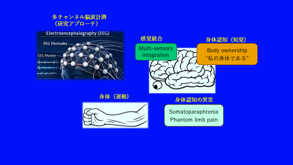
Neural Mechanisms of Bodily Self-Consciousness
To perform movements skillfully, we need to feel that our body belongs to us (i.e., the sense of body ownership) and that we ourselves are the agents who initiate and control our movements (i.e., the sense of agency). For healthy individuals, these bodily self-experiences are taken for granted. However, in various conditions?such as somatoparaphrenia after stroke or phantom limb pain following limb amputation?bodily self-consciousness can be impaired, often hindering subsequent recovery. Bodily self-consciousness is a new research field in which many aspects of the underlying neural mechanisms remain unclear. Our research aims to understand the neural mechanisms of bodily self-consciousness from a neuroscientific perspective and to develop novel neurorehabilitation approaches based on these insights. In this project, we seek to elucidate the neural mechanisms of bodily self-experiences using tools such as electroencephalography (EEG), transcranial magnetic stimulation (TMS), and virtual reality (VR).
Research equipment: multi-chanel electroencephalography, 3D motion capture system, liquid crystal apparatus for tachistoscopic occlusion, polygraph system, visuo-haptic stimulator, electrodermography
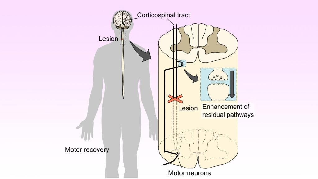
Development of Novel Therapies that Promote Motor Recovery Following Brain and Spinal Cord Injuries
For us to move our own body, motor commands from the brain must reach the spinal cord. The most well-known pathway for this is the corticospinal tract. However, the corticospinal tract can be damaged by compression or trauma, leading to impaired motor function or paralysis. On the other hand, animal studies (in monkeys) have shown that even when the corticospinal tract is damaged in the spinal cord, motor recovery can still occur if the pathway mediated by interneurons remains intact. However, in humans, who have a highly developed corticospinal tract, the neural connections of interneurons are thought to be relatively “weak.” Therefore, we hypothesize that effective motor recovery may be possible if we can artificially enhance the efficiency of interneurons, and we are developing specific methodologies to achieve this. To this end, we are conducting research on human participants with and without neurological diseases using techniques such as motion analysis, electromyography, and transcranial magnetic stimulation. This project is being conducted in collaboration with the Department of Orthopedic Surgery and the Department of Medical Physiology at the Kyorin University School of Medicine.
Research equipment: 3D motion capture system, electromyography, transcranial magnetic stimulator, transcranial direct current stimulator, neuronavigation system, peripheral nerve and muscle stimulator
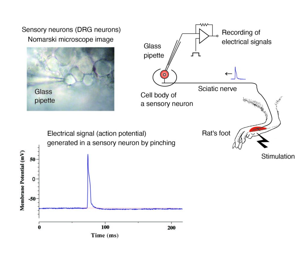
Mechanisms of Electrical Signal Encoding in Pain
The peripheral nerves that convey touch, temperature, pain, and itch are highly diversified. How do these sensory neurons detect and transduce diverse stimuli?such as touch, pinching, heat, cold, pain-inducing substances, and ischemia?into signals, thereby forming their corresponding sensory modalities (subtypes of sensory experiences)? To answer this classic question in physiology, we have independently developed the “In Vivo Patch-Clamp Recording of Sensory Neurons” and are investigating the mechanisms by which stimuli from both inside and outside the body are encoded into electrical impulses.
Research equipment: system for recording electrical signals from sensory nerves in anesthetized rats